简体中文
简体中文

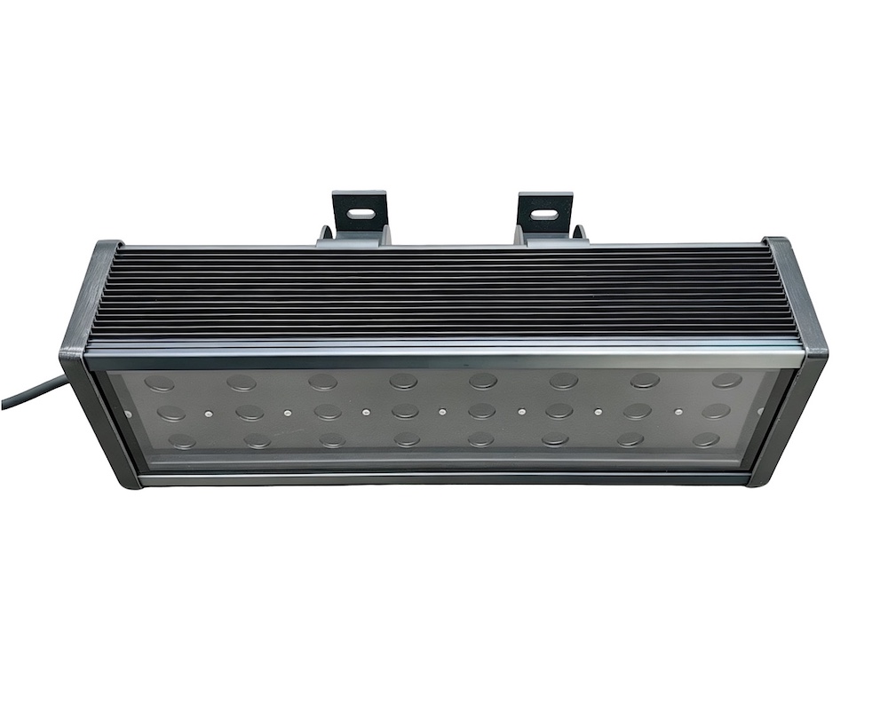
Researchers from Henan University optimized a protein fluorescence reporting system for cotton to accelerate somatic embryogenesis regeneration screening and functional gene labeling. The team employed a fluorescent protein excitation lamp LUYOR-3415RG with fluorescent filter sets for green and red fluorescence observation.
Abstract
Protein fluorescence reporting systems are of crucial importance to in-depth life science research, providing systematic labeling tools for visualization of microscopic biological activities in vivo and revolutionizing basic research. Cotton somatic cell regeneration efficiency is low, causing difficulty in cotton transformation. It is conducive to screening transgenic somatic embryo using the fluorescence reporting system.
However, available fluorescence labeling systems in cotton are currently limited. Too ptimize the fluorescence reporting system of cotton with an expanded range of available fluorescent proteins, we selected 11 fluorescent proteins covering red, green,yellow, and cyan fluorescence colors and expressed them in cotton. Besides mRuby2 and G3GFP, the other nine fluorescent proteins (mCherry, tdTomato, sfGFP, Clover, EYFP, YPet, mVenus, mCerulean, and ECFP) were stably and intensely expressed in transgenic callus and embryo, and inherited in different cotton organs derive from the screened embryo.
In addition, transgenic cotton expressing tdTomato appears pink under white light, not only for callus and embryo tissues but also various organs of mature plants, providing a visual marker in the cotton genetic transformation process, accelerating the evaluation of transgenic events.
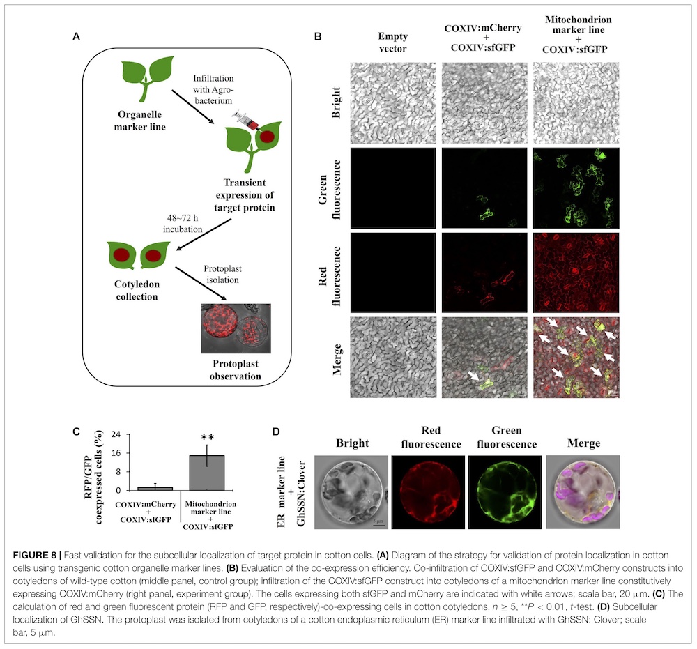
Further, we constructed transgenic cotton expressing mCherry-labeled organelle markers in vivo that cover seven specific subcellular compartments: plasma membrane, endoplasmic reticulum, tonoplast, mitochondrion, plastid, Golgi apparatus, and peroxisome. We also provide a simple and highly efficient strategy to quickly determine the subcellular localization of uncharacterized proteins in cotton cells using organelle markers.
Lastly, we built the first cotton stomatal fluorescence reporting system using stomata-specific expression promoters (ProKST1, ProGbSLSP, and ProGC1) to drive Clover expression. The optimized fluorescence labeling system for transgenic somatic embryo screening and functional gene labeling in this study offers the potential to accelerating somatic cell regeneration efficiency and the in vivo monitoring of diverse cellular processes in cotton.
Fluorescence Observation Using LUYOR-3415RG Fluorescent Protein Excitation Lamp
The fluorescence in embryonic callus, stamen, pistil, leaf, and seed tissues of transgenic cotton or tobacco epidermal cells was observed with a zoom-stereo microscope (OLYMPUS, Tokyo, Japan). The fluorescence in protoplast, stomatal cell, fiber, leaf epidermal cell, and root samples of transgenic cotton was observed with a confocal electron microscopy (Nikon, Tokyo,Japan).
Transgenic samples larger than 2 cm2 were observed using a hand-held LUYOR-3415RG fluorescent protein excitation lamp (LUYOR, Shanghai, China), and images were captured using an EOS 800D camera (Canon, Beijing, China) with a fluorescent filter set (LUYOR) for green and red fluorescence observation.
Green or yellow fluorescence was observed with a filter set for excitation at 488 nm and emission at 500–550 nm; red fluorescence was observed with a filter set for excitation at 561 nm and emission at 570–620 nm; cyan fluorescence was observed with a filter set for excitation at 405 nm and emission at 425– 475 nm.
FIGURE 1: Stable expression of red and green fluorescent proteins (RFPs and GFPs, respectively) in cotton callus and embryo tissues.
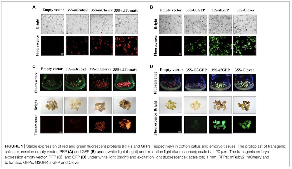
FIGURE 2: Red fluorescent protein (RFP) fluorescence in different tissues/organs of transgenic regenerated T0 plants.
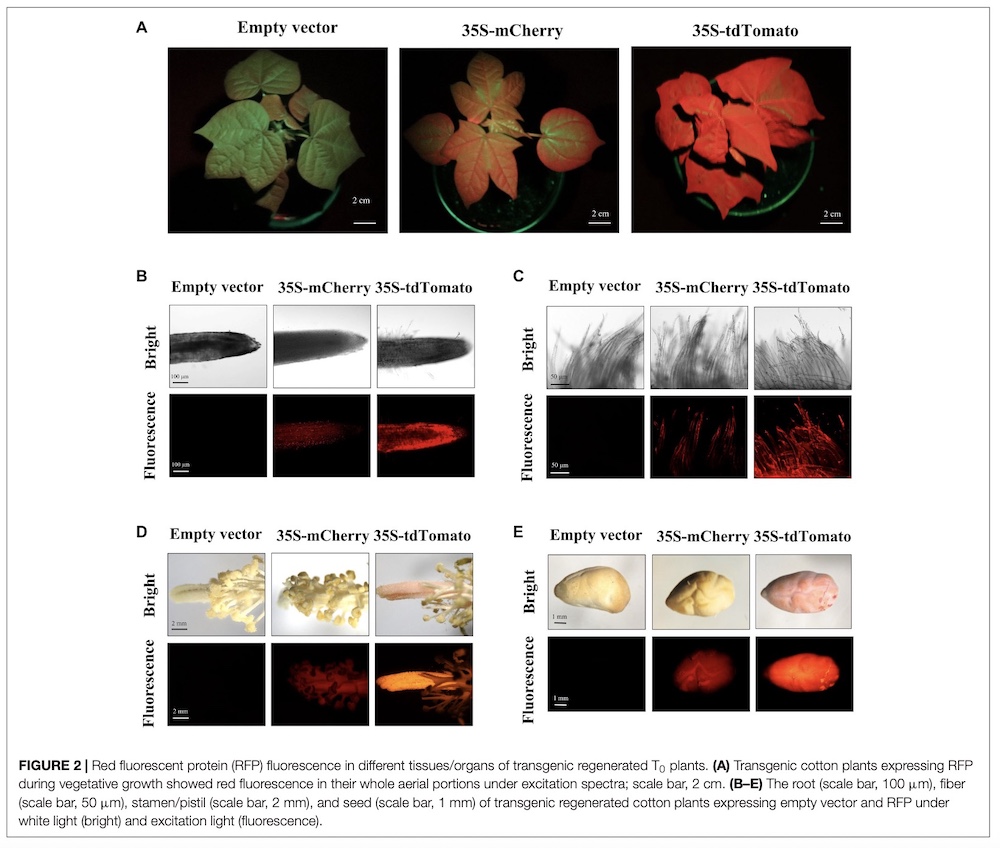
FIGURE 3: Green fluorescent protein (GFP) fluorescence in different tissues/organs of transgenic regenerated T0 plants.
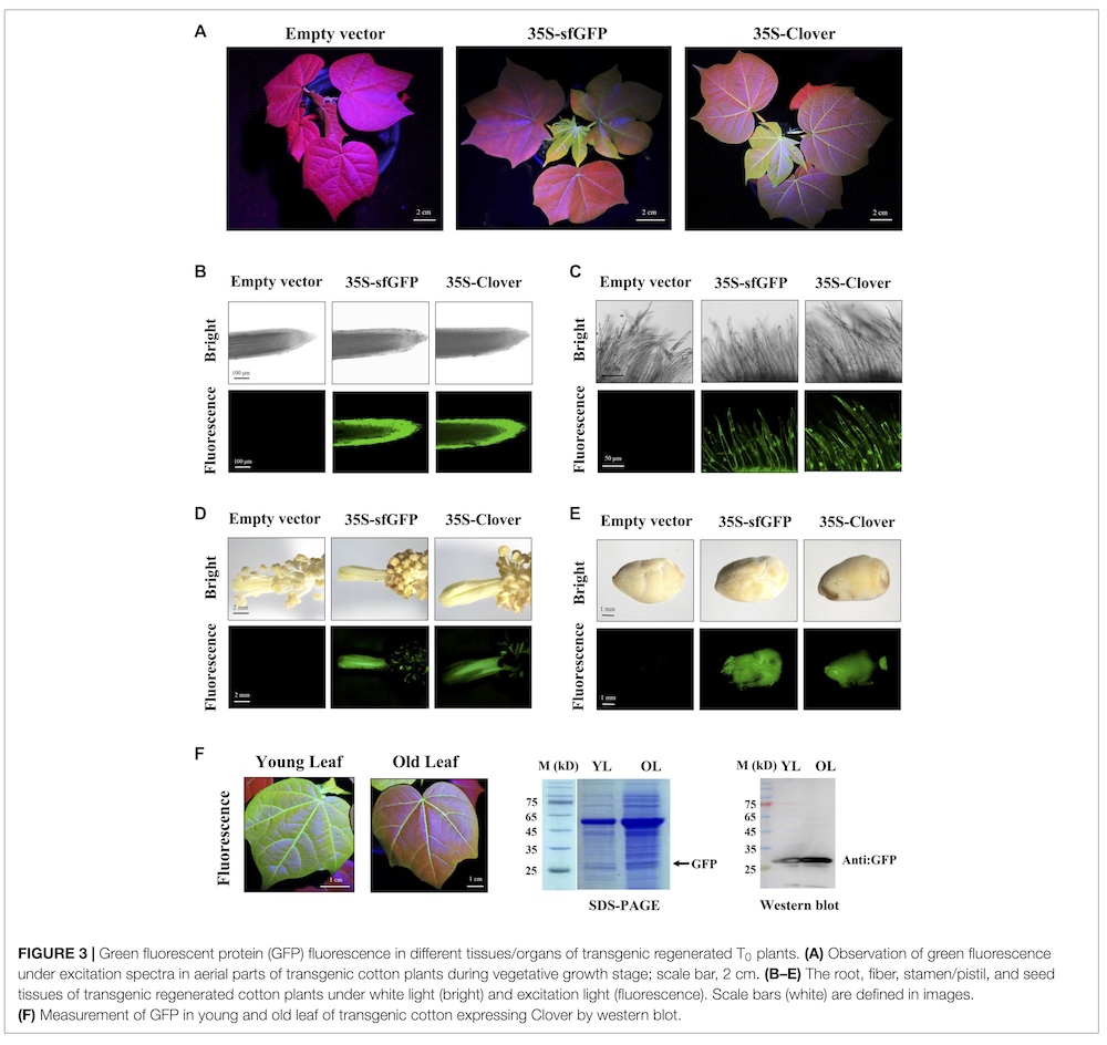
FIGURE 4: Yellow and cyan fluorescent protein (YFP and CFP, respectively) fluorescence in different tissues/organs of transgenic regenerated T0 plants.
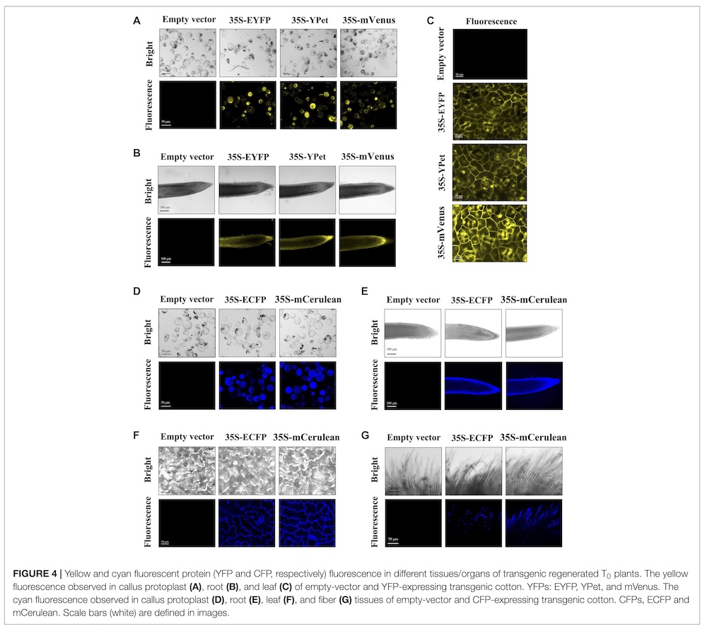
FIGURE 5: The fluorescence of T1 progeny in field conditions.
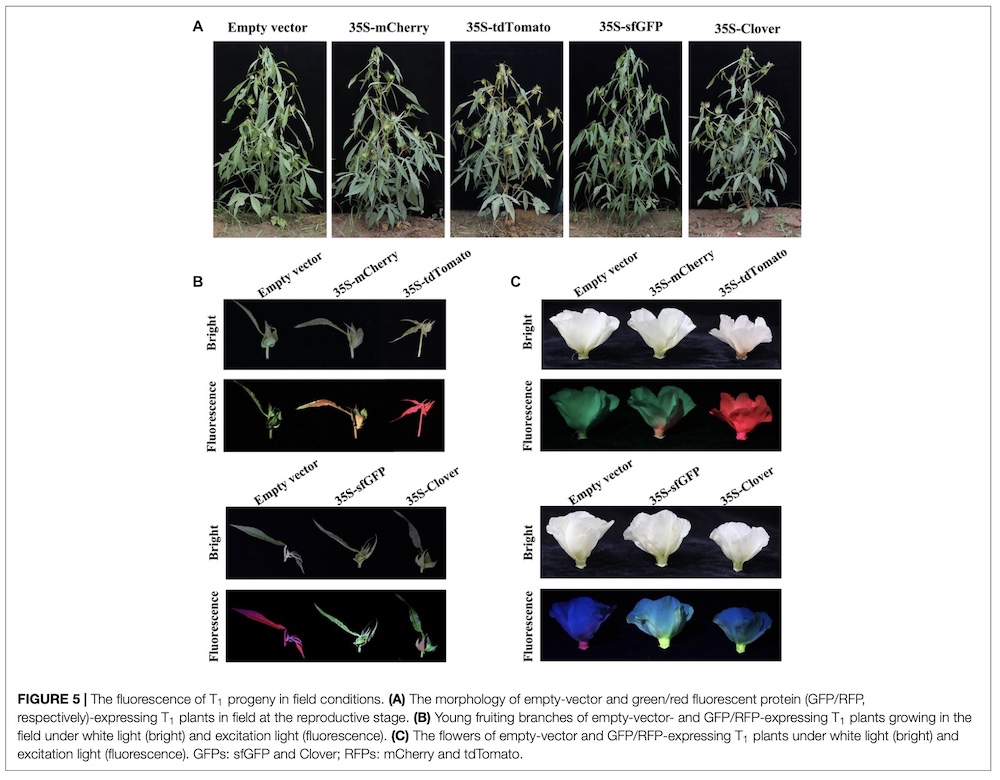
FIGURE 6: The tdTomato protein serves as a visual marker for successful transgenic events.
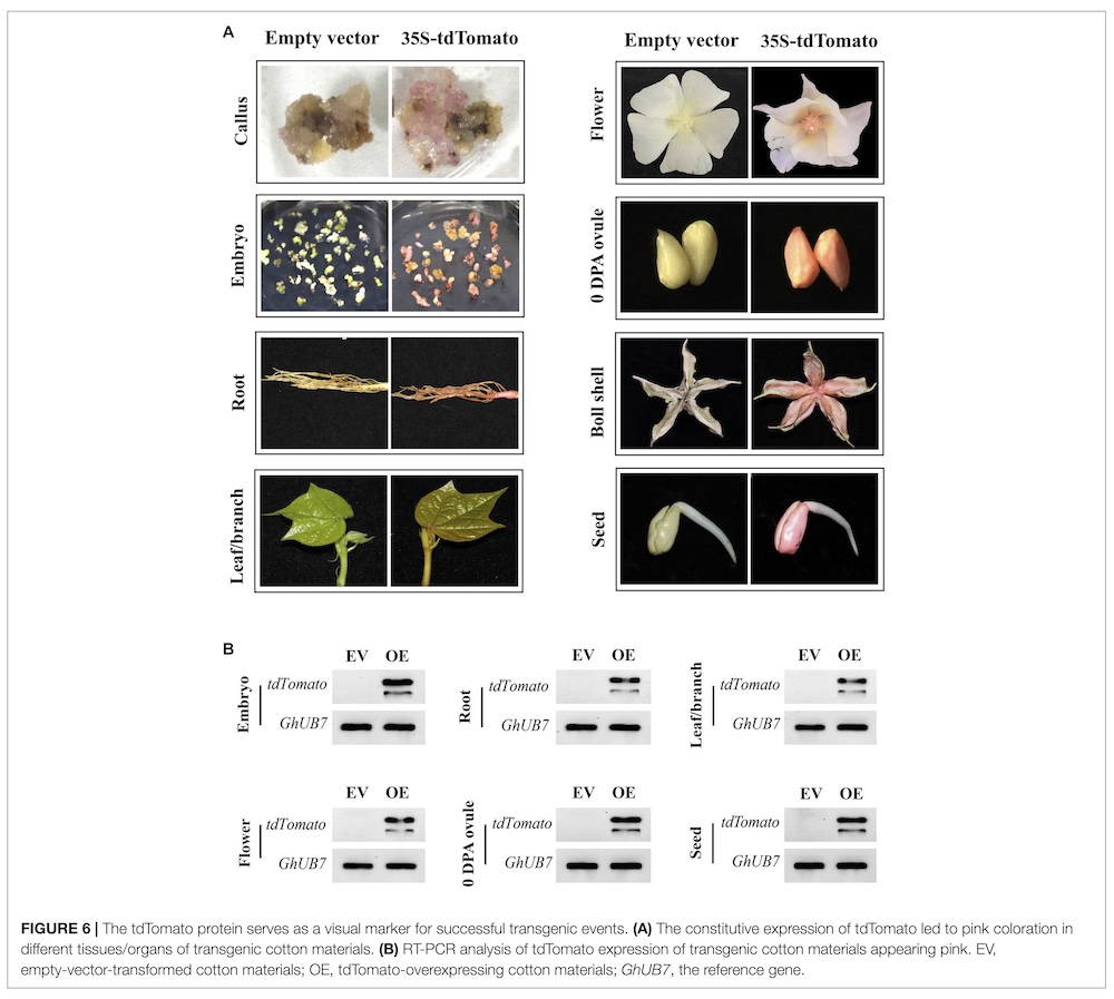
FIGURE 7: Transgenic cotton expressing in vivo organelle markers.
FIGURE 8: Fast validation for the subcellular localization of target protein in cotton cells.
Reference
Hu, G.-Y., Ma, J.-Y., Li, F., Zhao, J.-R., Xu, F.-C., Yang, W.-W., Yuan, M., Gao, W., & Long, L. Optimizing the protein fluorescence reporting system for somatic embryogenesis regeneration screening and visual labeling of functional genes in cotton. Front. Plant Sci. 12, 825212 (2022).